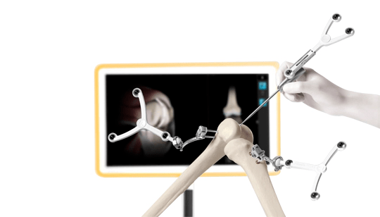
Knee Joint Preservation refers to the use of nonsurgical or surgical means to preserve a deteriorating joint in order to delay or avoid joint replacement surgery.
When cartilage deterioration due to osteoarthritis is causing persistent joint pain that interferes with your daily life, it is our goal to restore normal movement and alleviate pain to your joint – be it your shoulder, hip, or knee. Joint preservation refers to the use of nonsurgical or surgical means to preserve a deteriorating joint in order to delay or avoid joint replacement surgery. Every patient is different, so our specialists will customize your joint preservation strategy with you based on your individual situation, taking into account factors such as your age, expectations, level of joint dysfunction, and activity level.
When cartilage deterioration due to osteoarthritis is causing persistent joint pain that interferes with your daily life, it is our goal to restore normal movement and alleviate pain to your joint – be it your shoulder, hip, or knee. Joint preservation refers to the use of nonsurgical or surgical means to preserve a deteriorating joint in order to delay or avoid joint replacement surgery. Every patient is different, so our specialists will customize your joint preservation strategy with you based on your individual situation, taking into account factors such as your age, expectations, level of joint dysfunction, and activity level.


The knee could be a changed hinge joint, a kind of articulatio, that consists of 3 purposeful compartments: the patellofemoral articulation, consisting of the patella, or “kneecap”, and also the os sesamoideum groove on the front of the thighbone through that it slides; and also the medial and lateral tibiofemoral articulations linking the thighbone, or thigh bone, with the shin, the most bone of the lower leg.[6] The joint is bathed in synovia that is contained within the tissue layer referred to as the joint capsule. The posterolateral corner of the knee is a section that has recently been the topic of revived scrutiny and analysis.
The knee is that the largest joint and one amongst the foremost necessary joints within the body. It plays an important role in movement associated with carrying the weight in horizontal (running and walking) and vertical (jumping) directions.
At birth, the kneecap is simply shaped from animal tissue, and this may ossify (change to bone) between the ages of 3 and 5 years. as a result of it’s the most important bone within the anatomy, the ossification method takes considerably longer.
The main body part bodies of the thighbone square measure its lateral and medial condyles. These diverge slightly distally and posteriorly, with the condyle being wider ahead than at the rear whereas the condyle is of a lot of constant breadth. The radius of the condyles’ curvature in the sagittal plane becomes smaller toward the rear. This decreasing radius produces a series of involute midpoints (i.e. settled on a spiral). The ensuing series of cross axes allow the slippery and rolling motion within the flexing knee whereas making certain the collateral ligaments square measure sufficiently lax to allow the rotation related to the curvature of the condyle a couple of vertical axis.
The combine of leg bone condyles square measure separated by the intercondylar eminence composed of a lateral and a medial tubercle.
The patella also serves AN body part body, and its posterior surface is stated because the trochlea of the knee. It is inserted into the skinny anterior wall of the joint capsule. On its posterior surface may be a lateral and a medial body part surface,[10]:194 both of that communicate with the patellar surface which unites the 2 limb condyles on the anterior aspect of the bone’s distal finish.
Knee Joint Preservation techniques are used to repair damaged joints and delay the need for joint replacement surgery. Among the surgical Knee Joint Preservation procedures are:
- Knee osteotomy – this is a surgical procedure for patients who have osteoarthritis in just one of the knee compartments. It can help to relieve pain and increase movement in the knee.
- Cartilage transplant – cartilage grown in the laboratory is used to replace damaged cartilage or the bones are stimulated to promote cartilage growth.
- Microfracture – using arthroscopy, multiple holes are made in the bone, around 4mm apart. Bone marrow cells and blood then covers the area helping to promote the growth of new tissue.
- Autologous osteochondral transfer – this involves harvesting bone and cartilage from parts of the knee that bear less weight and transferring this to the damaged area.
- Autologous chondrocyte implantation – a small piece of articular cartilage is harvested from the patient’s knee and sent to the laboratory to be treated with enzymes to isolate the chondrocytes, which are cells that produce cartilage. These are multiplied and implanted into the patient’s body six to eight weeks later.
There are several non-surgical procedures for relieving pain, including:
- Corticosteroid injections
- Injections of hyaluronic acid
- Platelet-rich plasma injections
Although Knee Joint Preservation surgery has now become a routine procedure it is still a major operation that carries risks. These risks are higher for people who are overweight, as well as for smokers and people with other health conditions. Sometimes, delaying surgery may be advisable to give people the opportunity to quit smoking, lose weight or stabilise their health. Joint preservation surgery may be recommended as a way of repairing the damaged joint and postponing Knee Joint Preservation surgery for as long as possible.
In addition, prosthetic implants have a lifespan of around 15 to 20 years so younger patients who undergo Knee Joint Preservation surgery are more likely to need revision knee joint preservation, a surgical procedure to replace the worn-out implant. This type of surgery carries an increased risk of complications. Prosthetic implants may also be subject to loosening, stiffness and infection, which can also require surgery.
joint preservation surgery is used in patients with significant joint pain but who are not yet ready to undergo Knee Joint Preservation surgery. This may be because they are younger (and so more likely to need revision knee joint preservation), because they lead an active lifestyle or because they have very localised arthritis.
Knee Joint preservation surgery is carried out arthroscopically (using keyhole surgery).
During a knee osteotomy, a wedge of bone is removed from the upper shinbone or lower thighbone. This shifts the body’s weight off the damaged area of the knee joint and not the opposite side of the knee where cartilage remains intact. Metal plates and screws are used to hold the bones of the knee in their new position. In some cases, a graft of bone may be used to help the osteotomy to heal faster. After the procedure you will normally stay in hospital for one or two days during which time a physical therapist will recommend exercises to help to build strength and flexibility in your knee.
You will normally need crutches for four to six weeks after surgery to keep the weight off your knee as it heals. You may be able introduce low impact exercises such as walking and cycling as your joint heals.
Four to six months after joint preservation surgery you should experience a significant improvement in movement and joint strength. It will allow you to continue to living a normal life by reducing pain to a manageable level and increasing your mobility. You may still need joint replacement surgery but it will enable you to postpone the date of surgery
What is knee joint preservation surgery?
Knee joint preservation surgery is a new philosophy in the sphere of knee surgery, which aims to preserve the human knee for as long as possible. This has come about over the last three decades of joint replacement surgery, being the mainstay of treatment for degenerative knee disease.
We have found through research and a collection of outcomes that knee joint preservation surgery is successful cases and that following knee replacement, a further operation is required to re-do the knee replacement usually after around 10 to 15 years. As patients are developing knee joint degenerative problems at a younger age, we have developed different approaches to avoid the need for knee replacement surgery.
When is this treatment offered?
Knee Joint Preservation surgery is offered after a thorough assessment, including patient history, clinical examination, X-rays of the knee and long leg alignment views. The long leg alignment views allow the clinician to assess the patient’s mechanical loading access. This helps us to find out at which point through the knee the load is passing.
Humans have three different types of alignment. There is a neutral alignment, which is a straight leg, a varus alignment, which appears as bowed legs, and valgus alignment which appears as knocked knees.
If patients have bowed legs or knocked knees and subsequent to that have worn out one specific part of their knee (either the medial or lateral side of the joint) then the underlying pathology, which is the mechanical malalignment, can be corrected. This is called osteotomy surgery and can be combined with biological therapies, such as platelet-rich plasma injections to maintain the cartilage in the knee joint.
Will all knee joint preservation patients need replacement surgery eventually?
The results of joint preservation /osteotomy surgery are generally very good with 85% of patients not requiring joint replacement surgery after 10 years. This means that only 15% of those patients will go on to require a joint replacement operation.
The reason for this is that osteoarthritis is a progressive condition and despite the realignment of the leg the osteoarthritis can continue. More modern techniques, as well as adding biological therapies into the knee joint, can delay knee replacement surgery even further.
