Knee Joint Preservation refers to the use of nonsurgical or surgical means to preserve a deteriorating joint in order to delay or avoid joint replacement surgery. best knee joint preservation in chhindwara
When cartilage deterioration due to osteoarthritis is causing persistent joint pain that interferes with your daily life, it is our goal to restore normal movement and alleviate pain to your joint – be it your shoulder, hip, or knee. Joint preservation refers to the use of nonsurgical or surgical means to preserve a deteriorating joint in order to delay or avoid joint replacement surgery. Every patient is different, so our specialists will customize your joint preservation strategy with you based on your individual situation, taking into account factors such as your age, expectations, level of joint dysfunction, and activity level. best knee joint preservation in chhindwara
When cartilage deterioration due to osteoarthritis is causing persistent joint pain that interferes with your daily life, it is our goal to restore normal movement and alleviate pain to your joint – be it your shoulder, hip, or knee. Joint preservation refers to the use of nonsurgical or surgical means to preserve a deteriorating joint in order to delay or avoid joint replacement surgery. Every patient is different, so our specialists will customize your joint preservation strategy with you based on your individual situation, taking into account factors such as your age, expectations, level of joint dysfunction, and activity level. best knee joint preservation in chhindwara

The knee could be a changed hinge joint, a kind of articulatio, that consists of 3 purposeful compartments: the patellofemoral articulation, consisting of the patella, or “kneecap”, and also the os sesamoideum groove on the front of the thighbone through that it slides; and also the medial and lateral tibiofemoral articulations linking the thighbone, or thigh bone, with the shin, the most bone of the lower leg.[6] The joint is bathed in synovia that is contained within the tissue layer referred to as the joint capsule. The posterolateral corner of the knee is a section that has recently been the topic of revived scrutiny and analysis.
The knee is that the largest joint and one amongst the foremost necessary joints within the body. It plays an important role in movement associated with carrying the weight in horizontal (running and walking) and vertical (jumping) directions.
At birth, the kneecap is simply shaped from animal tissue, and this may ossify (change to bone) between the ages of 3 and 5 years. as a result of it’s the most important bone within the anatomy, the ossification method takes considerably longer.
The main body part bodies of the thighbone square measure its lateral and medial condyles. These diverge slightly distally and posteriorly, with the condyle being wider ahead than at the rear whereas the condyle is of a lot of constant breadth. The radius of the condyles’ curvature in the sagittal plane becomes smaller toward the rear. This decreasing radius produces a series of involute midpoints (i.e. settled on a spiral). The ensuing series of cross axes allow the slippery and rolling motion within the flexing knee whereas making certain the collateral ligaments square measure sufficiently lax to allow the rotation related to the curvature of the condyle a couple of vertical axis.
The combine of leg bone condyles square measure separated by the intercondylar eminence composed of a lateral and a medial tubercle.
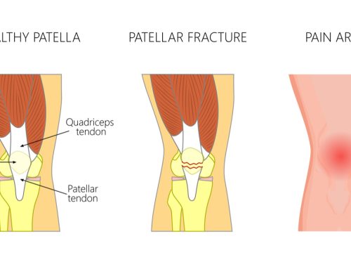
The patella also serves AN body part body, and its posterior surface is stated because the trochlea of the knee. It is inserted into the skinny anterior wall of the joint capsule. On its posterior surface may be a lateral and a medial body part surface,[10]:194 both of that communicate with the patellar surface which unites the 2 limb condyles on the anterior aspect of the bone’s distal finish.

